You Might Just Be Weird
The clinical significance of normal — and not so normal — anatomical variations
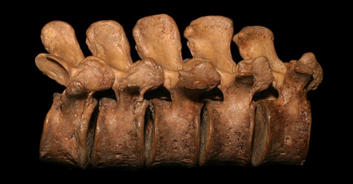
Note the significant differences in the size of the spinous processes of these lumbar vertebrae. This photo is from the collection of Paul and Suzee Grilley and is used with permission. They are all intended to “show the normal variation in human bones. None of them are pathological.”
Anatomy is not engineering — it’s wetter and sloppier. Even the best and most modern anatomical diagrams depict average anatomy only. Strange, wonderful, and problematic anatomical variations occur in humans all the time. My father, a US Navy corpsman (the navy equivalent of an army medic) has told me stories about the surprising number of minor and major birth defects he witnessed while working in a maternity ward in a Naval Hospital in Japan: webbings, large skin tags, extra digits, and so on. Here are several interesting examples that are also potentially clinically significant, especially in musculoskeletal medicine:
- Successful sprinters have unusually long heel bones (calcanei), which gives their calf muscles superior leverage. Bones shapes are amazingly variable, as shown in this bone photo gallery.1
- Dancers are particularly rife with unusual anatomy: ninety percent of a few dozen ballet dancers had some degree of hip dysplasia… a condition that affects only 1-5% of the general population.2 The same study identified a few more differences from non-dancer skeletons.
- About 1 in 10000 people have their organs arranged in a mirror image (situs inversus) — the liver is on the left, the heart on the right, and so on.
- Weird bones are normal, but the variability in the vertebrae3 and the pelvis4 has important implications for popular methods of diagnosing spinal joints being “out” or allegedly problematic pelvic tilts. I’ll elaborate on this one below.
- Surprisingly often, people have extra or missing ribs and vertebrae. The textbooks say that two floating ribs is standard (11th and 12th), but the truth is that roughly half the population, including yours truly, also has a 10th that floats free.5 This is based on very fresh data, and very few healthcare professionals are aware of it, but it has potentially significant clinical implications: due to their greater length and instability, floating 10ths probably sublux and twist more than 11ths and 12ths.
- Nerves in the heel, which might be related to frustrating persistent pain, are highly variable.6 Some people have one medial calcaneal nerve … some have three.
- Some folks have an extra knee bone, a second kneecap in back — the fabella instead of the patella — embedded in the tendon of the gastrocnemius muscle.7 How many people have this osseous oddity? About "20 to 87%" of people. Even the incidence is variable!8
- The size and shape of a notch in the top of the shoulder blade is quite variable, and nerve impingement is much more likely if you’ve got the wrong type of notch.9
- A Buford complex is a deformity of the glenoid labrum (rim of shoulder socket cartilage), a missing section in the 1-3 o’clock 🕑 position, plus a thickened, cord-like glenohumeral ligament. About 3% of individuals have this! Not exactly rare. Naturally, it can get mistaken for a rip. Radiopaedia.org: “There are a number of glenoid labral variants, whose importance is mainly due to the fact that the unwary may misinterpret them as pathology.”
- Radicular pain is the pain of an irritated nerve root, which causes symptoms in allegedly predictable and distinctive patterns that were first established over a century ago. When we started re-checking them decades later, surprise surprise, it turned out that things aren’t so simple.10 In a cadaver study in 2000, no two corpses’ nerve roots were quite alike. Although nerve roots themselves were quite consistent, they had a rat’s nest of random small branches connecting them, like the tangled roots of a tree.11
- Sometimes the sacrum is fused to the lowest lumbar vertebra by a bridge of bone, creating a “transition vertebra” — a vertebra that can’t decide if it’s lumbar or sacral. They are the most common congenital anomaly of the low back, found in a whopping 7% of the population (minimum)… and they probably do cause some trouble, the pesky little buggers.12 Or maybe not.13 It’s not totally clear!
- In about 6% of buttocks, the sciatic nerve and/or piriformis muscle get funky. The piriformis muscle may be split, and the sciatic may pass under, over, between, or even right through the muscle. And, yes, this can probably cause some trouble.14
- The vertebral artery in the neck is notoriously tortuous and variable: it can spiral and twist its way up the neck in configurations completely unnecessary for the delivery of blood. Unfortunately, sometimes it veers right into other structures, and presses on them with surprising intensity, relentless, throbbing …15
- Another abnormality related to the vertebral artery involves the holes in the vertebrae that it passes through: a “retrolenticular vertebral artery ring” is an abnormal bony bridge over the vertebral artery as it passes over the posterior arch of the atlas.16 People with this “feature” would suffer severe arterial compression during rotatory upper cervical manipulation, which is a fairly common chiropractic treatment, and a very dangerous possibility.
- Yet another neck item: there are some soft tissue connections between the spinal cord wrapping (dura mater) and the muscles of the upper neck — basically some rogue gristle. Exactly what’s connected and how tightly is unknown and likely inconsistent.17 The clinical implications are unclear, but they surely exist.18 CMBs probably explain why some people can flex their upper neck more comfortably than others, and they probably cause some headaches. Indeed, they may be a sneaky way that cervicogenic headaches work (cervicogenic = “from the neck”). Worse consequences are possible too: regular slight tugging on the spinal cord could really suck.19
- Os trigonum syndrome is an ankle affliction caused by an extra bit of bone (the os trigonum) in the joint, a minor anatomical variation caused by a developmental failure or a fracture. That little chunk of bone can get “jammed” in the complex mechanism of the joint, which is why it’s also charmingly called “the nutcracker-phenomenon.”
- Chinook headaches are the headaches people get from chinook winds, which are the warm, dry winter winds on the east side of the Rockies (AKA “Foehn Winds” and others around the world). They have a reputation for causing headaches, and Chinook winds do indeed seem to boost migraines.20 I expected that weirdness to be a total mystery, but was happily surprised to discover a decent hypothesis: the shape of the nose/throat plumbing is quite likely to explain chinook headaches!21 🤯 Specifically, some shapes may be prone to “locking the natural sinus ostia, thus preventing adequate pressure equilibrium.” While that doesn’t fully explain anything, of course, it’s a lot better than just “it’s weird, isn’t it?”

The “other kneecap”… the one in back! The fabulous fabella forms only in some lucky people.
The word ‘normal’ is probably an inappropriate word to apply to the human body.
Choosing Running Shoes: The Evidence Behind the Recommendations, Griffiths (SportsPodiatryInfo.co.uk)
Irregularity is to be expected in any biological form. Body parts are not interchangeable legos or Ikea furniture pieces made by factory molds. Wonkiness and asymmetry are part of the plan.
Playing With Movement, by Todd Hargrove, p. 169
The line between “normal variation” and “defect” is blurry.
For every visible, superficial oddity, there may be an invisible internal one. Surgeon Sherwin Nuland writes:
Of the many hundreds of appendectomies I have done during a career of thirty-five years, no two were the same. … Although the configurations of human innards do not vary nearly as much as do those of our outards (a word that exists in no dictionary but should), they nevertheless reveal unmistakable variations among individuals — and only surgeons ever find out about them.
See below for a longer excerpt from Nuland’s fantastic book.
And for every striking variation, there are probably a dozen minor ones. Any of them have the potential to be involved in musculoskeletal problems. In 2004, Hislop and Tierney wrote in the Journal of Science and Medicine in Sport:
… anatomical variations may be present, such as supernumerary [extra] muscles, thickened fascial bands or variant courses of nerves and blood vessels, which can themselves manifest as acute or chronic conditions that lead to significant morbidity or limitation of activity.
Hislop M, Tierney P. Anatomical variations within the deep posterior compartment of the leg and important clinical consequences. Journal of Science & Medicine in Sport. 2004;7:392–399.
For example, consider this story, reported by Costa et al. It concerns a cyst (which isn’t strictly an “anatomical variation” — it’s more of a straightforward abnormality, a “growth”). But the exact same thing could have happened with a cyst-like anatomical variation:
We present the case of a 28-year-old competitive runner with iliotibial band syndrome associated with a synovial cyst … open exploration revealed a large cyst beneath the ITB arising from the capsule of the knee proximal to the lateral meniscus. The cyst disappeared on extension. The preoperative MRI scan may have revealed the cyst, if it had been taken with the knee flexed.
Costa ML, Marshall T, Donell ST, Phillips H. Knee synovial cyst presenting as iliotibial band friction syndrome. Knee. 2004;11(3):247–248.
So the cyst basically created an atypical case of knee pain that defied diagnosis without surgical exploration. This person could easily have gone through a dozen doctors and therapists before finally getting some answers … and with no one really to blame. The body is just a weird place.

Most anatomical texts show only two floating ribs, just the 11th and 12th, which is considered “normal.” But this classic Gray’s Anatomy (plate 112), shows three floating ribs: the 10th, 11th, and 12th! As we now know, roughly half of people have 10th floaters (see Laswi). And 10th floaters are probably associated with some problems.
The X-files of therapy
Therapists and doctors will routinely make a great fuss over visible anatomical variations, often concocting elaborate treatment plans based on them.22 All the while, there could easily be invisible anatomical variations that are actually more important. (And bear in mind that there’s also a lot of overlap with functional variations.) It’s a classic and chronic clinical error: paying attention only to that which is easy to pay attention to, looking for your lost keys only where the light is good (“streelight effect”). It’s obviously foolish, and yet difficult to avoid. Out of sight really is out of mind.
The most implication of all this is:
- Invisible anatomical oddities are probably the cause of many stubborn and severe cases, especially pain with no obvious explanation, and probably makes some of them virtually impossible to recover from. Such cases often join the “X-files” of therapy, the unsolved clinical mysteries that every healthcare professional has seen too many of over the years.
Here are some other considerations:
- Recovery from some X-file cases is probably possible, but may explain why recovery is slow and difficult. Anyone struggling with a slow recovery should be (a little) reassured by this: there are perfectly good reasons why it can take much longer for some people. Slow progress doesn’t necessarily mean you’re doing it wrong.
- In general, injuries are more personal and idiosyncractic than we want to think. There’s a tendency for both patients and professionals to think of the common injuries as pretty well-defined animals, but the reality is that the lines between them blur — and it has a lot to do with anatomical variation.
- The “popular” biomechanical problems (i.e. pronating, short legs, spinal mis-alignment) are probably much less than half the story, and this should be considered before spending a lot of time or energy trying to fix them.
- Most invisible anatomical variations will never be detected or resolved. They are not just invisible, but in many cases virtually undiagnosable. They might be detectable in theory, but in practice never will be, because it will never make sense to go in there surgically to look — or to look as thoroughly as might be required.
Feeling for crookedness of the vertebrae and pelvis
Manual therapists (massage, chiro, etc) are often taught to rely on feeling the tips of vertebrae to judge the condition of the spine. Not all, but many.23 That’s certainly what I learned. In my first couple years as a baby Registered Massage Therapist, circa 2001,24 I would earnestly try to determine what spinal joints were “out”25 based on the position of spinal bone bumps.
And that makes me roll my eyes so hard these days. 🙄 Because now I know that you just cannot count on bones being nice and tidy and regular. They are no way to judge the position of vertebrae!
For instance, a 2022 study by Fausone et al. measured the lumbar spinous processes in 16 cadavers and found them to be quite variable in length.26
People just have funny bone shapes, and that has been shown in many ways over the years; this is more specific confirmation in the case of the lumbar spine. So it’s doubtful that anyone can find a “clinical positional fault of a vertebra through palpatory exam.”
A small statement that, if true, is a major bummer for the many professionals who have been trying to do exactly that, or relying on it to some extent.
This newer lumbar study is identical in spirit to a 2008 paper (Preece et al.) about pelvic bone shapes, which were also all the heck over the place, and which undoubtedly makes assessment of pelvic tilts highly unreliable.27 (And which I also used to do, arg, cringe.) Not that it’s even worth trying, crookedness being so generally overrated as a factor in pain and injury.
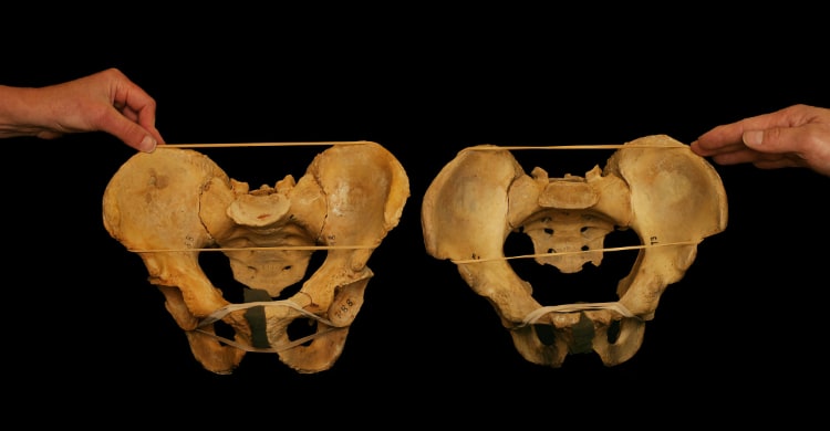
Another image from the collection of PaulGrilley.com, this one showing that bony landmarks of the pelvis are inherently irregular. It is not possible to diagnose any pelvic assymetry based on the apparent positions of the bones.
Did you find this article useful? Interesting? Maybe notice how there’s not much content like this on the Internet? That’s because it’s crazy hard to make it pay. Please support (very) independent science journalism with a donation. See the donation page for more information & options.
About Paul Ingraham

I am a science writer in Vancouver, Canada. I was a Registered Massage Therapist for a decade and the assistant editor of ScienceBasedMedicine.org for several years. I’ve had many injuries as a runner and ultimate player, and I’ve been a chronic pain patient myself since 2015. Full bio. See you on Facebook or Twitter., or subscribe:
Related Reading
- “Bone Photo Gallery — Bones,” Paul Grilley, PaulGrilley.com.
- The mysteries within: a surgeon explores myth, medicine, and the human body (book), by Sherwin B Nuland. Amazon.com ❐
- “Individual Differences: The Most Important Consideration for Your Fitness Results that Science Doesn’t Tell You,” James Krieger and Bret Contreras, Bretcontreras.com. Fascinating, readable tour of the many surprising (genetic) differences in the how people respond to the same diet and exercise.
What’s new in this article?
Five updates have been logged for this article since publication (2009). All PainScience.com updates are logged to show a long term commitment to quality, accuracy, and currency. more
Like good footnotes, update logging sets PainScience.com apart from most other health websites and blogs. It’s fine print, but important fine print, in the same spirit of transparency as the editing history available for Wikipedia pages.
I log any change to articles that might be of interest to a keen reader. Complete update logging started in 2016. Prior to that, I only logged major updates for the most popular and controversial articles.
See the What’s New? page for updates to all recent site updates.
2024 — Minor improvements.
2024 — Added an item about variants of the shape of nasal passages and sinuses being a plausible explanation for the phenomenon of chinook headaches.
2022 — Added a couple good citations about vertebrae and pelvic bone shapes, with some good images, and a new section: “Feeling for crookedness of the vertebra and pelvis.”
2019 — Added nerve root compression symptom pattern variability, which has an at least partly anatomical basis.
2018 — Added os trigonum syndrome; added syndactyly; plus some editing and reorganizing. This article is gradually evolving into something more than just a fun a cabinet of curiosities, as I continue to add clinically significant examples of anatomical variation.
2009 — Publication.
Appendix: a surgeon’s inside view of anatomical variation

The following excerpt is from the first chapter of Sherwin Nuland’s superb book, The Mysteries within: A surgeon explores myth, medicine, and the human body, “The Stomach: A Little Boy’s Big Secret.” I could not ask for a better passage to explain how strange and diverse “normal” anatomy can be:
No matter how often a surgeon performs the same operation, it is different each time. Any operating room nurse can tell you that. The sequential precision of predictable steps so exactingly depicted in manuals of surgical technique resembles the real thing about as much as a diagram of human anatomy looks like a human being.
To take something simple: Of the many hundreds of appendectomies I have done during a career of thirty-five years, no two were the same. Even such a straightforward operative procedure for a straightforward disease, divisible into a short series of straightforward technical maneuvers that were standardized almost one hundred years ago, done by an operator of long experience — even under such circumstances every case is a novelty. And some of those novelties can be daunting tests of skill and confidence.
On bedside rounds one day during the period of my training, the senior attending surgeon, a man highly respected for his dexterity and judgment, was asked to name the half-dozen most difficult operative cases he had encountered in his career. After a moment’s thought, he replied that three of them had been appendectomies. His answer surprised no one in the group of interns and residents who were crowded around him. In the few years of our embryonic surgical experience, every one of us had already seen enough to appreciate what he meant.
Although the configurations of human innards do not vary nearly as much as do those of our outards (a word that exists in no dictionary but should), they nevertheless reveal unmistakable variations among individuals — and only surgeons ever find out about them. The way in which an organ is attached by ligaments and folds of tissue to its surroundings, for example, is in general predictable, and yet just enough personal difference occurs that an operator never knows beforehand whether the viscus he is approaching will come up easily into his seeking hands or require deep dissection to free it. Friend appendix, in fact, epitomizes this kind of anatomical uncertainty. Being attached to the large intestine only at one narrow end of its wormlike body, the appendix is free to turn upward toward the liver, downward toward the pelvis, sideward toward the center of the abdomen, forward toward the abdominal wall, or even retrocecal, which means it has tucked itself up behind the bowel into a hidden location. The appendix may be as short as a stumpy inch or as willowy as five or six times that length. There is no telling where its tip may be found. Other organs, though not as variable, have unpredictabilities of their own.
And then there is the problem of fat. The copiousness of the cushions of fatty tissue lying between internal structures depends in general on an individual’s station along the spectrum between leanness and obesity. Thin people are a great deal easier to operate upon than are the chubbies, who hide vital structures deep within thick, greasy blankets of adiposity. Among those concealed vital structures are blood vessels, which have an obnoxious tendency to make uncharted course changes now and then, obstinately refusing to reach their destination via the route assigned to them by anatomy books. Lying in wait for the unwary, or perhaps lurking within a fatty bolster, an unanticipated artery or vein — and nerves are known to do this too — can affect the entire plan of a surgeon’s work, and sometimes its outcome.
Beyond even these considerations, the occasional occurrence of a congenital variation of structure must be taken into account. The operating team always has to be on guard for such an abnormality, especially because it may involve blood vessels or the slender ducts that carry secretions and other vital fluids to their destinations. Some of these inborn irregularities can present major challenges, or at least major surprises. From time to time, for example, one or another viscus or a part of it must be sought in an area of the body where it seems not to belong. I am not at all unique among surgeons to have removed thyroid tissue from the chest, found the right colon on the left side of the abdomen, and taken an ovary or appendix out of a hernia bulging into the uppermost part of the thigh.
Just to promote Nuland’s excellent book, I will continue the excerpt a little longer, even though the topic changes. He goes on to explain many more things that make every operation unique, even the common ones. But then he introduces an exotic case …
I have been referring here to operations done with relative frequency. For the reasons given or others, some cases will be so unusual that they stand out in a surgeon’s mind for the rest of his life. But in addition to that list, there exists a special category within even less commonly done procedures — these are the real rarae aves. By this I mean the one-and-onlies. These are the operations of such a unique type that the members of the team will regale one another with their details when they meet at reunions or conventions decades later, even in farflung parts of the world. Some of these procedures are firsts, or at least firsts in a given hospital — the first organ transplant, the first use of the heart-lung machine, the first video-controlled gallbladder operation — but some are memorable because no member of the team has ever seen their like before or since. Like all surgeons, I have a few of those once-in-a-lifetime adventures tucked away in the back of my mind, ready to be pulled out and relived at a moment’s notice.
One of them involves the stomach.
My patient had been an independent citizen for all of six weeks, the first two of which were spent in the preemie unit …
It’s a great story!
Notes
- PaulGrilley.com [Internet]. Grilley P. Bone Photo Gallery — Bones; [cited 18 Aug 14]. PainSci Bibliography 55592 ❐
- Harris JD, Gerrie BJ, Varner KE, Lintner DM, McCulloch PC. Radiographic Prevalence of Dysplasia, Cam, and Pincer Deformities in Elite Ballet. Am J Sports Med. 2016 Jan;44(1):20–7. PubMed 26324404 ❐
- Fausone D, Doherty D, Creighton D, Roach VA. Variations of spinous and transverse process length in the human lumbar spine. J Man Manip Ther. 2022 May:1–5. PubMed 35604056 ❐
- Preece SJ, Willan P, Nester CJ, et al. Variation in pelvic morphology may prevent the identification of anterior pelvic tilt. J Man Manip Ther. 2008;16(2):113–7. PubMed 19119397 ❐ PainSci Bibliography 52062 ❐
- Laswi M, Lesperance R, Kaye A, et al. Redefining the costal margin: A pilot study. J Trauma Acute Care Surg. 2022 Dec;93(6):762–766. PubMed 36121266 ❐
- Govsa F, Bilge O, Ozer MA. Variations in the origin of the medial and inferior calcaneal nerves. Arch Orthop Trauma Surg. 2006 Jan;126(1):6–14. PubMed 16333630 ❐
- Driessen A, Balke M, Offerhaus C, et al. The fabella syndrome - a rare cause of posterolateral knee pain: a review of the literature and two case reports. BMC Musculoskelet Disord. 2014;15:100. PubMed 24666711 ❐ PainSci Bibliography 54189 ❐
- Driessen: "The presence of the fabella in humans varies widely and is reported in the literature to range from 20% to 87%."
- Sangam MR, Sarada Devi SS, Krupadanam K, Anasuya K. A study on the morphology of the suprascapular notch and its distance from the glenoid cavity. Journal of Clinical and Diagnostic Research. 2013 Feb;7(2):189–92. PubMed 23542385 ❐ PainSci Bibliography 54576 ❐
- Rainville J, Laxer E, Keel J, et al. Exploration of sensory impairments associated with C6 and C7 radiculopathies. Spine J. 2016 Jan;16(1):49–54. PubMed 26253986 ❐
The dermatome patterns most professionals are familiar with were established many decades ago, and were not studied much again until the 21st Century. This study carefully checked the exact location of symptoms in 120 patients with suspected radiculopathy (symptoms in a dermatomal pattern, caused by nerve root compression). Perhaps unsurprisingly, they found that the dermatomal patterns were not as precise as the old maps would lead us to believe, and exhibit significant overlap, “to the extent that caution should be exercised when predicting compression of either the C6 or C7 nerve roots based on locations of impaired sensation.”
- Tanaka N, Fujimoto Y, An HS, Ikuta Y, Yasuda M. The anatomic relation among the nerve roots, intervertebral foramina, and intervertebral discs of the cervical spine. Spine (Phila Pa 1976). 2000 Feb;25(3):286–91. PubMed 10703098 ❐
- Sekharappa V, Amritanand R, Krishnan V, David KS. Lumbosacral transition vertebra: prevalence and its significance. Asian Spine Journal. 2014 Feb;8(1):51–8. PubMed 24596605 ❐ PainSci Bibliography 54202 ❐
Sometimes the sacrum is fused to the lowest lumbar vertebra: a lumbosacral transition vertebra. “LSTV is the most common congenital anomaly of the lumbosacral spine.” In about a thousand patients studied, it was about twice as common in patients who had sought spinal surgery as it was in patients with no spinal complaint (about 14-16% of patients, instead of 8%). The study also identified a “definite causal relationship” with degeneration of the disc above the LSTV.
- Apazidis A, Ricart PA, Diefenbach CM, Spivak JM. The prevalence of transitional vertebrae in the lumbar spine. Spine J. 2011 Sep;11(9):858–62. PubMed 21951610 ❐
- Natsis K, Totlis T, Konstantinidis GA, et al. Anatomical variations between the sciatic nerve and the piriformis muscle: a contribution to surgical anatomy in piriformis syndrome. Surg Radiol Anat. 2014 Apr;36(3):273–80. PubMed 23900507 ❐
This dissection study of 275 dead buttocks found that 6.4% of them had variations of sciatic nerve and piriformis muscle anatomy, with considerable variety in the variation. They found several different arrangements, and concluded: “Some rare, unclassified variations of the sciatic nerve should be expected during surgical intervention of the region.” Prepare to be surprised, surgeons!
All of these differences are potentially clinically significant, probably especially in the cases where the nerve (or part of it) passes right through the muscle. For a couple case studies, see Arooj 2014 and Kraus 2015.
- Kim HS, Lee JH, Cheh G, Lee SH. Cervical radiculopathy caused by vertebral artery loop formation: a case report and review of the literature. J Korean Neurosurg Soc. 2010 Nov;48(5):465–8. PubMed 21286489 ❐ PainSci Bibliography 54199 ❐
- Rao SR, Swamy G, Vasudha TK, Rao TR. Unusual foramen on the posterior arch of atlas. Journal of Science. 2015; 5(12):1165-1167.
- Palomeque-Del-Cerro L, Arráez-Aybar LA, Rodríguez-Blanco C, et al. A Systematic Review of the Soft-Tissue Connections Between Neck Muscles and Dura Mater: The Myodural Bridge. Spine (Phila Pa 1976). 2017 Jan;42(1):49–54. PubMed 27116115 ❐
This review of 26 studies found “strong evidence” and concluded that it “proved” that there are connections between some suboccipital muscles and the dura mater, while there is “limited evidence” and “controversy” about others. They conclude: “There is a continuity of soft tissue between the cervical musculature and the cervical dura mater.”
- Enix DE, Scali F, Pontell ME. The cervical myodural bridge, a review of literature and clinical implications. J Can Chiropr Assoc. 2014 Jun;58(2):184–92. PubMed 24932022 ❐ PainSci Bibliography 53727 ❐
- Consider the evidence that chronic intermittent spinal cord irritation is strongly associated with fibromyalgia: see Holman.
- Cooke LJ, Rose MS, Becker WJ. Chinook winds and migraine headache. Neurology. 2000 Jan;54(2):302–7. PubMed 10668687 ❐
- Rudmik L, Muzychuk A, Oddone Paolucci E, Mechor B. Chinook wind barosinusitis: an anatomic evaluation. Am J Rhinol Allergy. 2009;23(6):e14–6. PubMed 19769801 ❐
- Expensive orthotics for your feet, tedious exercises to get you “aligned,” and so on.
- Obviously, many chiropractors and osteopaths are trained better and know better. Not all, however! And I have known all too many who do in fact have this simplistic view … harmonizing nicely with many other simplistic and dubious beliefs that are disturbingly prevalent in those professions.
- I was a Registered Massage Therapist with a busy practice in Vancouver, Canada, from 2000–2010. I quit the profession in disgust before I could be “fired” for being critical of pseudoscience and alternative medicine. I changed my professional focus to science journalism and anti-quackery activism, writing mostly about massage-adjacent topics in the early years, but then spreading out into anything relevant to any kind of chronic pain or injury rehab — an effectively infinite list of topics. Even fifteen years after my last hands-on work, I still think of myself as a massage therapist. That said, I don’t exactly feel like an “insider” either, and I’ve branched out a lot in that time. See my bio.
- The “outness” of spinal bone joints is mostly the chiropractic concept of a spinal joint “subluxation,” which can be defined in many ways, from reasonable to nonsensical. See Spinal Subluxation: Can your spine be out of alignment? Chiropractic’s big idea has been misleading patients for more than a century.
- Fausone 2022, op. cit.
- Preece 2008, op. cit.